Thursday, November 12, 2015

Heart Dissection Lab

Q1: What is the purpose of the pericardium?
The pericardium is a double sac of serous membrane that secretes a fluid to lubricate the heart and reduce friction. It also protects the heart.


Q2: Observe the blood vessels connecting to the heart.  How do arteries differ from veins in their structure?
Veins have thinner walls than arteries and are also one way valves (prevent back flow of blood). Arteries carry blood away from the heart to the tissue while veins carry blood to the heart from the tissue. Veins store the most blood volume in the body. Arteries are elastic and contractile.


Q3: Place your finger inside the auricle.  What function do you think the auricle serves?
The auricle feels rough when we placed our finger inside the auricle. Auricle increases blood capacity and volume in the atrium and also collect blood.


Q4: Observe the external structures of the atria and ventricles.  What differences do you observe?
The right atria is the upper chamber that receives oxygen deprived blood. The right ventricle a lower chamber that discharges blood. The left atria is the upper chamber that receives oxygen rich blood. The left ventricle is a lower chamber that has the thickest wall. In structure, the ventricle has thicker walls and takes up more space.

Q5: Find and Describe the following structures in the heart:
1. Coronary Sinus: a wide venous channel that receives blood from the coronary veins and empires into the right atrium of the heart.
2. Inferior Vena Cava: (carries blood from lower body) a large vein and empties into the right atrium of the heart. The superior vena cava carries blood from upper body.
3. Tricuspid Valve: Located between the right atrium and right ventricle; it prevents back flow of blood into the atrium


Q6: Draw a picture of the tricuspid valve, including chordate tendinae and the papillary muscle.
Picture:


Q7: Why is the “anchoring” of the heart valves by the chordate tendinae and the papillary muscle important to heart function?
After the right ventricle contracts, the blood pressure pushes on the tricuspid valve, which stops the blood flow to the right atria. The job of chordae tendinae is to make sure the side edges of the valve don’t get pushed into the atria. It is important because it stops the valve from pushing blood in the wrong direction.

Q8: Using pictures and/or words describe what you see (bicuspid valve)
Picture:

The probe in this picture points to the bicuspid valve, which is between the left atrium and ventricle


Q9: What is the function of the semi-lunar valves?
The Semilunar valves prevent arterial blood from re-entering the heart. Two types are the pulmonary semilunar valve and the aortic semilunar valve.


Q10: Valvular heart disease is when one of more heart valves does not work properly.  Improperly functioning heart valves can lead to regurgitation, which is the backflow of blood through a leaky valve.  Ultimately this can lead to congestive heart failure, a condition that can be life threatening.
If the valve disease occurs on the right side of the heart, it results in swelling in the feet and ankles.  Why might this happen?
The ventricles are not strong enough to pump blood against gravity from your toes and feet. Blood is not able to function normally and back flow occurs.
If the valve disease occurs on the left side of the heart, what complications would you expect to see?
If the valve disease occurs on the left side of the heart, not enough blood would be pumped. Therefore, swelling would occur.

Q11: Using pictures and/or words describe what you see (left/right coronary arteries, left semilunar valve (3 cusps), chord tendinae of bicuspid valve, and pillar muscle of the bicuspid valve)
Picture of left ventricle:


a. Entrance to the left/right coronary arteries: The coronary arteries supply blood to the heart.
b. Left (aortic) semilunar valve (3 cusps): The aortic semilunar valves prevent arterial blood from re-entering the blood.
c. Chordae Tendinae of the Bicuspid Valve: Chordae Tendinae are tendons attached to cusps of valves.
d. Papillary Muscle of the Bicuspid Valve: They provide "muscle power" and they pull on the chordae tendinae to open/close valve.

Q12: Describe how the left and right sides of the heart differ from each other.
The right side of the heart pumps deoxygenated blood to the lungs. The left side of the heart pumps oxygenated blood to the aorta and out to the rest of  the body. Also, the right side of the heart has thinner walls while the left side of the heart has thicker walls.


Q13: Draw and label all structures visible in the interior of the cross-section.
Picture:


Tuesday, November 10, 2015

Unit 3 Reflection

This Unit was about the circulatory system, the respiratory system, and cardiovascular diseases such as stroke and heart disease. The circulatory system transports materials needed by the cells and removes waste materials from the cells. The respiratory system works with the circulatory system to exchange gas. The cardiovascular system works by the pulmonary circuit (O2 deprived) and the systemic circuit (O2 rich). Blood first enters the right atrium from the inferior and superior vena cava. It then passes through the tricuspid valve and enters the right ventricle. It passes through the pulmonary semilunar valve to the pulmonary trunk to the pulmonary arteries and out to the lungs. Lungs then exchange carbon dioxide for oxygen. In the systemic circuit, pulmonary vein returns blood to heart via the left atrium. It passes through the bicuspid (mitral) valve and enters left ventricle. Lastly, passes through aortic semilunar valve into aorta and out to body. Veins carry blood to the heart from the tissues while arteries carry
blood away from the heart to the tissue. 
Cardiovascular health refers to the health of the heart and blood vessels. Lack of exercise, being overweight, smoking, metabolic syndrome, diabetes, high blood pressure (caused by atherosclerosis), and high cholesterol all cause cardiovascular disease. To promote cardiovascular health, people should control their blood pressure. 40-90% of all stroke victims had high blood pressure before their stroke. To maintain healthy blood vessels, people should exercise, maintain a healthy weight and diet, control blood pressure and cholesterol, prevent atherosclerosis, prevent diabetes, don’t smoke, and minimize stress. I want to learn more about Lung Cancer, Chronic Bronchitis, Asthma, Cystic Fibrosis, Sinusitis, Tuberculosis, Pleurisy, Influenza, etc. because those chronic pulmonary diseases seem very interesting to me. My aunt had lung cancer so I would love to learn more about it and how it can affect someone’s life. I wonder if there has been any medications to prevent strokes kind of like a vaccine.
My weaknesses in this unit were learning the diagram of the heart. I had a lot of trouble understanding the different parts of the heart even though I understood the functions of all the major valves and blood vessels. I enjoyed the heart dissection even though it was very difficult to locate all the parts of the heart. Working in a group helped me with this challenge. This unit was very interesting to me because some of my family members have had heart attacks (they never recovered fully after) so I finally understand what could have caused the disease. I also had trouble studying for this test because I tried many methods to try to understand all the material. I studied with Nimisha the day before to make sure we both understand the topics. I am still having trouble studying because even though I put a lot of effort into my studying, it doesn’t seem to help because I still don’t do good on the tests. My strengths in this unit was being able to identify the functions of the different blood vessels and the different diseases (causes and effects). 
My health goals (from Unit 2 Reflection) were to eat more fruits and to sleep more. I have been eating more fruits; however, my amount of sleep has remained the same because of tennis and work from school. It is hard to balance out both my tennis practice and homework. For the remainder of the semester, I hope to spend more time understanding the pictures because they are very helpful in understanding the unit.

Pictures:


For the video above, we made one mistake toward the end of the video. After the left ventricle, we said it goes through bicuspid vale but it actually goes through the aortic semilunar valve into aorta and out to the body.
Picture one is a picture of the heart we drew on the street. Picture two is a picture of the heart during dissection. Picture three is a selfie we took with the heart at the beginning.

Tuesday, October 13, 2015

Measuring Blood Pressure Lab

From my data, I learned that my systole was 120 and my diastole was 75 for test one. For test two, I learned that my systole was 140 and my diastole was 70. The two tests showed somewhat different results mostly likely because we did them so close to each other due to time. A typical blood pressure is 120/80 mm Hg. The first number represents the pressure when the heart contracts and is called the systolic blood pressure. The second number represents the pressure when the heart relaxes and it called the diastolic blood pressure.

This is my data for heart rates and blood pressure.

To measure heart rate, you need a stethoscope and your own two fingers to measure the pulse rate (radial and carotid). You use a stethoscope only when you find the pulse of your actual heart. Carotid arteries are located on your neck while radial arteries are located on your wrists. To measure blood pressure, you use an instrument called a sphygmomanometer. The sphygmomanometer contains an air bulb, valve, cuff, a pressure gage, etc.

Using your thumb to measure pulse is not ideal because there are large vessels located in the thumb that have their own pulse so it can interfere with the pulse you are trying to find.

Using a blood pressure cuff (sphygmomanometer) is simple once you understand the procedure. First, you wrap the cuff around the person's upper arm, making sure the cuff is located above the crease of the person's elbow. Make sure to put on a stethoscope so you can hear the pulse later on in the lab. Inflate the cuff gently while watching the pressure gauge. Keep inflating until the person's pulse disappears. When you reach about 150 mmHg, open the valve on the air pump slightly, making sure not too much air is released. Listen to the stethoscope carefully until you hear the heart beat start again. This is the systolic blood pressure (write it down). The sounds should continue to become louder. Write down when you hear the sound for the last time. This is called the diastolic blood pressure.

This is a picture of the stethoscope and sphygmomanometer we used.


Wednesday, October 7, 2015

Unit 2 Reflection

Health is the measure of our body's efficiency and over-all well being. The 5 pillars of health are very important to live a healthy life, which include stress, exercise, social, sleep, and nutrition. All of these pillars of health are very important and people should not focus on just a couple, they should focus on all of them. Being Healthy also corresponds to having healthy hormones. The "big 4" hormones are Insulin, Leptin, Cortisol, and Glucagon. Insulin is anabolic hormones secreted by alpha cells of the pancreas while Glucagon is the opposite: catabolic hormones secreted by beta cells of the pancreas. When insulin, leptin, glucagon, and cortisol are doing their job properly, you are physically healthy.After studying Unit 2 Health, I think I am healthy because I always follow the five pillars of health. Sleep is very important to me and I try to get as much as possible. I eat healthy and stay away from hydrogenated oils. I try not to get too stressed out because that can negatively affect my health. I play tennis every day and I also play on the school tennis team. Lastly, I spend a lot of times with friends. However, I don't think overall people at Saratoga High School are healthy mostly because they are not getting the necessary amount of sleep and they get too stressed out from school. Students procrastinate which makes them more likely to be sleep deprived. To promote greater health and wellness on campus, I think students should be given less strenuous homework assignments and students should try not to procrastinate. 

I learned a lot from the health unit. I did not know that stress and sleep can affect your overall well being. I also learned a lot from the Falcon Market because a lot of the foods I thought were healthy turned out to be very fatty. For example, some types of peanut butter contain hydrogenated oil, which I thought was very interesting because hydrogenated oil (transfat) is very bad for you and should be avoided as much as possible. I also learned a lot about the different types of hormones and the effect they have on the body. The themes and essential understandings of this unit was what is health, what is needed to maximize your nutrition for optimal health and disease prevention, how does exercise enhance health, why is sleep important, what are the physiological affects of stress, and why are social connections good for your health? I still don't fully understand all the functions of the hormones, such as leptin and cortisol because they are very complex. To improve on the next unit, I will focus more in class and try to stay more organized with all my worksheets. 

This unit relates to my life because my grandma got a very severe diabetes and I never fully understood why. However, now i know that diabetes is common with people who eat an excess about of sugars and unhealthy things. This relates to one of the five pillars of health: nutrition. To prevent from getting diabetes or other illnesses, people need to eat good food, including omega 3. 

Goal Sheet: My health goals are to eat more fruits and vegetables and to sleep more. The actions I will take is to pay more attention to the foods I eat and to not procrastinate so I can finish my homework earlier. A healthy life is important to me because I want to be healthy and I don't want to die from any health related reasons. I will know when I have reached my goal when I have eaten more vegetables and I sleep earlier. 
Images relating to blog post: (sleep, good nutrition, and exercise)


Saturday, October 3, 2015

Monday Wellness Reflection

Link to Google Presentation:

For our Monday Wellness Project, Cassi and I chose stretching because it is a really interesting topic and I feel that a lot of athletes don’t stretch because they think it is a waste of time. However, I wanted to inform the class that stretching does help and it should be done before and after exercise for many benefits that were discussed in the presentation. From our research on stretching, we learned that muscles need to be well circulated and healthy. A normal person spends most of the day in the same position. If people avoid stretching, their muscles will continue to fatigue. As a result of not stretching, people decrease their circulation and flexibility. As an athlete myself, I found this research very fascinating because before this presentation, I would rarely stretch before practice because I always thought that stretching was a waste of time. However, this presentation taught me that stretching is beneficial and now, I stretch before and after practice and games. Our Monday Wellness project has really inspired me to stretch myself.
The most interesting things that we learned while preparing for the presentation was Brad Walker’s research that explains that the current research on stretching is flawed. As we were looking through research, we noticed that a lot of the research done on stretching was done in the same way: two groups, one stretching and the other not stretching. This led us to Brad Walker’s article that talks about the flawed research because stretching shows effects after a long period of time. Also, we found that changing stretches too often, called “bouncing,” is not beneficial. This fact was very interesting to me because usually when I stretch, I would change my stretches often to get it over with, but now I know that doing that is not effective and shows no beneficial results. 
Stretching is important for health and wellness because it increases flexibility, increases range of motion, improves circulation, promotes better posture, etc. Stretching is very important and athletes should not just skip stretching because it is very beneficial. I think stretching should be enforced more in sports because it is very useful and ultimately helps people’s overall wellness. 
Overall, I would give our presentation a high 8 or low 9 out of ten because even though we spent a lot of time preparing for our presentation, we put too many videos in our presentation. This was not effective; instead, we should have just talked about the major points in the videos. I think that we should have explained more of the anatomy of the different stretches so students can understand how the major muscle groups work during stretching. We should have made the presentation more interactive so students would not get bored during our presentation. Also, I think that our information was very helpful and I hope people now understand that they should spend more time stretching. I feel that a lot of students don’t do stretches properly, mostly because they are too lazy, so I hoped that the stretches we did during class showed them the most effective ways to stretch in order to get the full effects of the stretch.


Wednesday, September 16, 2015

What is Health?


Health is very important to me. I believe health is a measure of our mental, social, and physical well being and condition. Good health is something that everyone wants in their life. In order to achieve good health, it is important to understand the 5 pillars of health, which are nutrition, exercise, sleep, stress, and social. I think the nutrition pillar is the strongest because good nutrition helps you grow and lead a healthy life style. If you eat bad food, you have a higher change of developing obesity and diabetes. I think I need the most improvement on the sleep pillar because I usually don't get enough sleep every day due to homework. I want to learn more about the causes of good health and the ways in which different people can become healthy.


Tuesday, September 8, 2015

Unit 1 Reflection

This unit was supposed to be an Intro to Anatomy and Physiology. We learned about the different anatomical terms, the four macro molecules, and the four different tissue types. The themes and essential understandings our Biology: cell bio and molecules, Anatomy and Physiology directional terms, and histology. I learned a lot about the different tissue types and the four macro-molecules. The labs helped me get a visual of the four tissue types, so I could understand how their form fits their function. I understand everything that we learned this chapter, but naming the different tissue types is still difficult for me to do. To improve in the next unit, I will make flashcards on the important materials so it can help me remember the information better. My aunt damaged her epithelial tissue and I didn't fully understand what could happen as a result of the injury. However, now I know that as a result of tissue damage, inflammation occurs which causes swelling, warmth, pain, and redness. These conditions are a result of mast cell activation which releases vasodiliators such as histamine.

3 Pictures from this Unit:

Picture from where I sit. At the beginning of each period, we do the do-now and look at the homework. This whiteboard also has the essentials and our agenda for the day.


Lab notebook and computer I use when I do blog posts at school.

Monday, September 7, 2015

Tissue Lab

This lab helped me learn about the different tissues and their structures. It was really interesting to see the different types of tissue under the microscope: epithelial, connective, muscular, and nervous tissue. After we looked at each tissue type, it's easier to understand their functions because form fits function and we could relate the tissues to the other types and look at their differences. This lab helped me understand the lecture notes more because in the lecture, we learned the different parts of the tissue and the functions. By observing the structure, its easier to understand how the tissue works in relation to the rest of the body. 

The differences between the different tissue types were obvious after looking at them through a microscope. For example, the connective tissue was tightly/ loosely backed with a bunch of tiny cells. Here is a picture of an epithelial cell from the microscope:


Here is a picture of the different types of connective tissue:


Here is a picture of the different nervous tissues:


Lastly, here is a picture I drew of the Human Skeletal Muscle (Muscle Tissue):

As you can see from the pictures, Epithelial Tissue looked like continuous sheets. The different shapes, such as simple squamous and stratified cuboidal, help account for the various functions of epithelial cells, like protection, absorption, secretion, and ion transport. The shape of the connective tissue was mainly tiny little fibers densely or loosely packed together. This shape helps fit the function of connective tissues, which is to bind together, support, and strengthen other body tissues. The shape of muscle tissues was either cylindrical (skeletal), branched (cardiac) or spindle shaped (smooth). The shape helps fit the function of each of the three muscle types.


Monday, August 31, 2015

Cell Specialization

Epithelial Cells

Epithelial Cells line the cavities of the body and cover the body. Epithelial cells come in a variety of shapes, including squamous epithelial, cuboidal epithelial, and culumnar epithelial. In addition to these shapes, epithelial cells can be described as being simple or stratified. Simple contains one layer while stratified contains multiple. (EC) Our skin is made up of epithelial cells.

Here are the different types of epithelial cells.


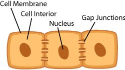
Below is the structure of an epithelial cell.  It differs from eukaryotic cells because it only contains a cell membrane, cell interior, and nucleus that a eukaryotic cell has. Epithelial cells have gap junctions, but eukaryotic cells don't.

gap junctions


Epithelial Cells are very complex cells and many functions that help the body function properly. For example, they help transport nutrients from one place in the body to the other. They also help guard against microbes. They are found in the lungs, skin, airway, and digestive tract. They also secrete the enzymes in the stomach. Moreover, all the functions of epithelial cells are protection, absorption, secretion, transportation, and filtration. (ET) Epithelial cells are classified as epithelial tissues. The cells functions depend on where they are found in the body. According to AboutHealth , "epithelial cells represent our first line of defense against pathogens that can enter out bodies through our airways, digestive tracts, and bloodstreams."


Tuesday, August 25, 2015

Sweetness Lab


In this lab, my teammates and I tasted the various types of carbohydrates: sucrose, glucose, fructose, galactose, maltose, lactose, starch, and cellulose. The relationship between taste and structure is that as the number of rings increases, the amount of sweetness in the carbohydrate decreases. I can relate some of these sugars to the foods I have eaten. For example, cellulose is in plants and we eat vegetables. It makes sense that cellulose is not sweet because vegetables, which contain cellulose, are not sweet. On the other hand, Fructose is very sweet. We can relate fructose to fruits because fruits contain fructose. Fruits are sweet and therefore, fructose is also sweet. Humans taste sweetness differently and therefore rank the various carbohydrates differently. Taste buds make up the papillae. According to Kids Health, "Taste buds are sensory organs that are found in your tongue and allow you to experience tastes that are sweet, salty, sour, and bitter." (Kids Health) People taste sweetness because when you chew food, chemicals are released that allow the taste buds to gain flavor and taste for the foods you eat. Different tasters rank the sweetness of the same samples differently because everyone has different tastes. According to ILoveBacteria, everyone has four taste buds which are sour, salty, bitter, and sweet. People who have more taste buds generally "are more sensitive to strong flavors" (ILB); they are called super tasters. In this lab, students ranked the different carbohydrates differently because of the degree of sweetness different people can taste and because everyone has different tastes.

This is a picture of the different carbohydrates we used during the lab. Each table group got one of each sugars and we were able to taste all of  them.